No products in the cart.

 Français
 Deutsch
 Italiano
 Português
 Español
 简体中文
简体中文Kundalini Yoga Intervention Increases Hippocampal Volume in Older Adults:
A Pilot Randomized Controlled Trial
Marim Ibrahim1, Joseph Therriault2, Vasavan P Nair3, Elena Dikaios4, Pedro Rosa-Neto5, Ishan C Walpola6, Soham Rej1, Michael Lifshitz7
1 Geri-PARTy Research Group, Jewish General Hospital; Department of Psychiatry, McGill University, Montreal, QC, Canada
2 Translational Neuroimaging Laboratory, McGill University Research Centre for Studies in Aging, Alzheimer’s Disease Research Unit, Douglas Research Institute, Le Centre Intégré Universitaire de santé et de Services Sociaux (CIUSSS) de l’Ouest-de-l’Île-de-Montréal; Department of Neurology and Neurosurgery, Psychiatry and Pharmacology and Therapeutics, McGill University, Montreal, Canada
3 Department of Psychiatry, McGill University; Douglas Mental Health University Institute, Montreal, QC, Canada
4 Department of Educational and Counselling Psychology, McGill University, Montreal, QC, Canada
5 Translational Neuroimaging Laboratory, McGill University Research Centre for Studies in Aging, Alzheimer’s Disease Research Unit, Douglas Research Institute, Le Centre Intégré Universitaire de santé et de Services Sociaux (CIUSSS) de l’Ouest-de-l’Île-de-Montréal; Department of Neurology and Neurosurgery, Psychiatry and Pharmacology and Therapeutics, McGill University; Montreal Neurological Institute, Montreal, Canada
6 Brain and Mind Centre and School of Medical Sciences, Faculty of Medicine, University of Sydney, Sydney, Australia
7 Department of Psychiatry, McGill University; Lady Davis Institute for Medical Research, Jewish General Hospital, Montreal, QC, Canada
Click here for correspondence address and email
| Date of Submission | 08-Feb-2022 |
| Date of Decision | 24-Jun-2022 |
| Date of Acceptance | 06-Jul-2022 |
| Date of Web Publication | 5-Sep-2022 |
Abstract
Background: Among a rapidly aging population, there is increased need for neuroprotective interventions promoting healthy neurological aging. Mind-body interventions, such as Kundalini yoga, are actively being explored as accessible means to encourage healthy aging. However, little remains known about the neurobiological effects of Kundalini yoga. Aims: This pilot randomized-controlled trial (RCT) examined the potential neuroprotective effects of Kundalini yoga in older adults. Methods: We conducted an RCT with 11 healthy meditation-naïve older adults. Participants were randomized to a Kundalini yoga or psychoeducation intervention. Structural magnetic resonance imaging data were obtained at baseline and 12-week follow-up. The primary outcome measure was gray matter volume of the bilateral hippocampi and bilateral posterior cingulate cortex. Results: We found significant right hippocampal volume increases specific to the Kundalini yoga group (P = 0.034, ηp2 = 0.408). Conclusions: These findings provide initial neurobiological support for the neuroprotective effects of Kundalini yoga.
Keywords: Gray matter volume, Kundalini yoga, mind-body intervention, neuroimaging, neurological aging
| How to cite this article: Ibrahim M, Therriault J, Nair VP, Dikaios E, Rosa-Neto P, Walpola IC, Rej S, Lifshitz M. Kundalini Yoga Intervention Increases Hippocampal Volume in Older Adults: A Pilot Randomized Controlled Trial. Int J Yoga 2022;15:158-62 |
| How to cite this URL: Ibrahim M, Therriault J, Nair VP, Dikaios E, Rosa-Neto P, Walpola IC, Rej S, Lifshitz M. Kundalini Yoga Intervention Increases Hippocampal Volume in Older Adults: A Pilot Randomized Controlled Trial. Int J Yoga [serial online] 2022 [cited 2022 Sep 12];15:158-62. Available from: https://www.ijoy.org.in/text.asp?2022/15/2/158/355600 |
Introduction
Given our rapidly aging population, there is an urgent need to explore protective interventions against age-related cognitive decline. Yoga interventions have been found to be feasible, safe, and effective in preventing cognitive decline and were found to display potential neuroprotective effects.[1],[2] Kundalini yoga integrates meditation, yoga postures, and breathing exercises (pranayama). It has been reported to promote healthy neurological aging. For instance, a 12-week Kundalini yoga program was shown to improve executive functioning and memory in older adults (n = 81) with mild cognitive impairment (MCI).[3] Another study found that an 8-week Kirtan Kriya meditation program (derived from Kundalini yoga) improved mood and decreased anxiety in older adult participants (n = 15) with memory loss.[4] The clinical and neurological benefits of Kundalini yoga appear promising.
However, the neurobiological effects of Kundalini yoga remain understudied, with no published studies examining structural neurobiology in healthy older adults. Thus, here we report results from a pilot randomized-controlled trial (RCT) examining the neurobiological effects of a 12-week Kundalini yoga intervention, compared to a 10-week psychoeducation intervention, among healthy older adults. In particular, we examined the effects of Kundalini yoga on gray matter volume (GMV) of the hippocampus and posterior cingulate cortex (PCC), two brain regions whose atrophy is associated with cognitive decline in neurotypical aging and early Alzheimer’s disease.[5]
Methods
Participants
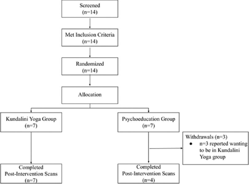
Fourteen participants were randomized with seven participants allocated to Kundalini yoga and 7 to psychoeducation [Figure 1]. Three participants withdrew from the psychoeducation intervention, stating they would have preferred to be in the yoga group. Eleven participants completed the study (n = 7 yoga, n = 4 psychoeducation).
Participants were aged 62–74 (M = 66.4, standard deviation [SD] = 3.1) with Mini-Mental State Exam (MMSE) scores between 28 and 30 (M = 29.7, SD = 0.6). In the yoga group, participants were aged 62–74 (M = 66.86, SD = 3.761), 57.1% were male and 42.9% reported a medical condition (e.g., high cholesterol, history of major depression, urinary incontinence, and high blood pressure which was being controlled) and taking medication (e.g., blood pressure medication). In the psychoeducation group, participants were aged 64–67 years (M = 65.50, SD = 1.291), 50.0% were male and 25.0% reported a medical condition (prior myocardial infarction and high blood pressure which was being controlled) and taking blood pressure medication. No significant baseline differences were found between groups for age, MMSE scores, or GMV for ROIs (Ps > 0.35). We found that all scans were unremarkable (Schentens scores ≤1) after rating images for white matter hyperintensities and atrophy of the hippocampal formation.
Participants were recruited through newspaper advertisements and from the Douglas Mental Health University Institute (Montréal, Canada). Participants were prescreened to ensure they were eligible for the study based on the inclusion and exclusion criteria. This screening process included the administration of the MMSE to assess cognitive functioning before participation.[6] We excluded participants who had prior experience with meditation, regularly consumed alcohol or tobacco, had magnetic resonance imaging (MRI) contraindications, diabetes, cardiovascular or respiratory illness, or any active psychiatric/neurological illness.
Written informed consent was obtained from all participants. The study protocol was approved by the research and ethics office of the Centre Intégré Universitaire de Santé et de Services Sociaux de l’Ouest-de-l’Île-de-Montréal (CIUSSS-ODIM)/Douglas Institute and was in accordance with the Helsinki Declaration of 1975.
Intervention groups
Participants were randomized 1:1 to a Kundalini yoga intervention or psychoeducation intervention. Participants were matched across groups for gender and age. Sessions were led by trained interventionists at the Douglas Mental Health University Institute. The Kundalini yoga intervention lasted for 12 weeks, and the psychoeducation intervention lasted for 10 weeks. The discrepancy in program length was the result of scheduling complications in which the interventionist leading the psychoeducation group was unable to complete a 12-week duration. Each group had one session per week which lasted 2 hours. Interventions were conducted bilingually in French and English.
The Kundalini yoga intervention was customized for participant safety and included a mixture of postures, pranayama, and meditation. In addition, participants were encouraged to practice at home for 30 min per day.
The psychoeducation intervention consisted of a psychoeducation program in which participants were taught about memory and healthy aging. Each of the weekly sessions included learning about prospective memory, working memory, and executive functions such as problem-solving and decision-making. As in the Kundalini Yoga intervention, participants in the psychoeducation group were encouraged to complete homework for 30 min per day.
Structural magnetic resonance imaging acquisition
A structural MRI scan was obtained before and after participation in the intervention. A structural MRI scan was obtained before and after participation in the intervention. We used a T1-weighted sequence. Images were acquired on a 3T Siemens Magnetom using a standard head coil. A volumetric magnetization prepared rapid gradient echo MRI (TR: 2300 ms, TE: 2.98 ms) sequence was employed to obtain a high-resolution T1-weighted anatomical image of the entire brain (9 degree flip angle, sagittal orientation, 1 mm × 1 mm in-plane resolution of 1 mm slab thickness). A parallel acquisition technique of GRAPPA was used.
Data processing and analysis
Structural MRI data were obtained at baseline and 12 weeks. We calculated GMV in four predetermined regions of interest (left and right hippocampus and left and right PCC) using the standard preprocessing pipeline and voxel-based morphometry toolbox in SPM12.[7] Regions of interest were defined based on the Desikan-Killiany-Tourville atlas.[8] The DARTEL algorithm was used to spatially normalize scans. We spatially registered and segmented scans into gray matter, white matter, and cerebrospinal fluid using the tissue probability maps in SPM12. Total intracranial volume was calculated, and data were smoothed using an 8 mm full width at half maximum Gaussian kernel. The final voxel size was 1.5 × 1.5 × 1.5 mm.
For each region of interest, we conducted a two-way repeated-measures ANOVA to examine the effects of condition (Kundalini yoga versus psychoeducation) and time (baseline vs. postintervention) on volume using JASP (https://jasp-stats.org/). An alpha of 0.05 was used for statistical tests. Levene’s test for equality of variance was used to ensure the assumption of homogeneity was met for each region of interest. All participants who completed the study were included in the data analysis.
Results
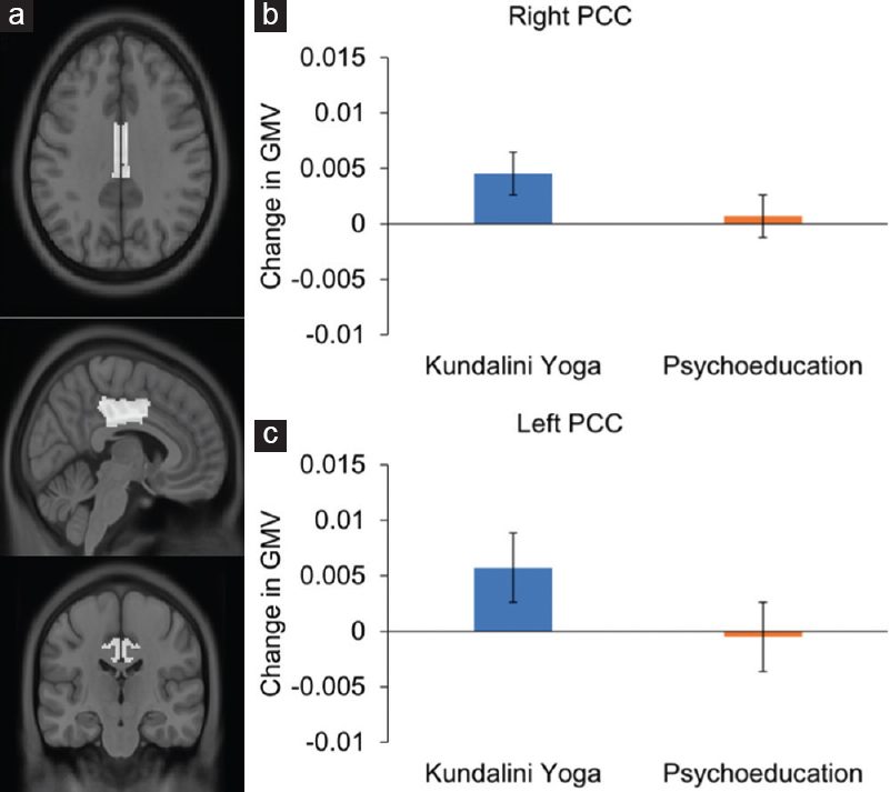
A group-by-time ANOVA yielded a significant interaction effect on right hippocampal volume (F (1,9) = 6.210, P = 0.034, ηp2 = 0.408). In the left hippocampus, this interaction effect was not statistically significant (F (1,9) = 4.747, P = 0.058). No significant changes were found in the PCC. Results are outlined in [Table 1] and [Figure 2] and [Figure 3].
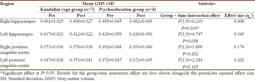

Scheffe’s test was used for post hoc analysis to decompose the significant interaction on volume of the right hippocampus. These post hoc tests yielded no significant differences between the cells. To further explore the data, we also conducted two simple contrasts that we felt were most relevant using a t-test (comparing pre- to post-intervention volume within each intervention type). For these two contrasts, we applied Bonferroni correction to adjust the alpha to 0.025 (critical alpha 0.05 divided by 2 tests). Results from these analyses showed an increase in volume of the right hippocampus from pre- to post-intervention in the Kundalini yoga group (t (6) = 3.109, P = 0.021), but not the psychoeducation group.
Discussion
To our knowledge, this is the first RCT using neuroimaging to investigate Kundalini yoga as a protective intervention against neurological aging in healthy older adults. We found that participants in the 12-week Kundalini yoga intervention showed significant increases in right hippocampal volume. Notably, the only other structural neuroimaging study of Kundalini yoga found no significant effect in hippocampal volume; however, that study compared Kundalini yoga to memory enhancement training and focused on participants with MCI.[9] These factors may have overshadowed the effect of Kundalini yoga and may explain why our results are more positive, corresponding with neuroimaging findings from other forms of yoga that similarly showed increases in hippocampal volume.[10]
Age-related atrophy of the hippocampus has been linked to age-related declines in short-term memory.[11] These neurotypical atrophies are often exacerbated in Alzheimer’s disease, with hippocampal atrophy acting as a marker for the early stages of Alzheimer’s.[12] The right hippocampus in particular seems to be relevant to cognitive decline. For example, one study found that right hippocampal GMV correlated to scores on the MMSE and Montreal Cognitive Assessment.[13] A previous study reported that atrophy of the right hippocampus appears to be greater and occurs earlier than the left hippocampus, with right hippocampal atrophy possibly preceding a diagnosis of Alzheimer’s years in advance.[14] According to the National Institute on Aging and Alzheimer’s Association, the ideal time to intervene is during the early pre-clinical phase of Alzheimer’s.[15] Thus, our preliminary finding that Kundalini yoga increases right hippocampal volume should encourage further research examining this intervention as a way to promote healthy neurological aging.
This pilot study had two important limitations due to our logistical constraints: the small sample size (n = 11) and the discrepancy between intervention durations (12 weeks for the Kundalini yoga intervention compared to 10 weeks for the psychoeducation intervention). While promising, our results would need to be confirmed in a larger trial with more stringent experimental controls.
Concluding remarks
We examined the neurobiological effects of Kundalini yoga in a pilot neuroimaging RCT with healthy meditation-naïve older adults. We found a significant increase in volume of the right hippocampus after participation in the 12-week Kundalini yoga intervention, but not in the psychoeducation group. While preliminary, these findings encourage future full-scale trials to assess the potential of Kundalini yoga as a neuroprotective intervention.
Ethical statement
Written informed consent was obtained from all participants. The study protocol has been approved by the research and ethics office of CIUSSS-ODIM/Douglas Institute. ClinicalTrials.gov: NCT04726072.
Acknowledgment
Marim Ibrahim and Joseph Therriault are co-first authors and Soham Rej and Michael Lifshitz are co-senior authors in this paper.
Financial support and sponsorship
The study was funded by the Kripalu Institute and the Mind and Life Institute. Soham Rej receives salary support from the Fonds de Recherche du Québec Santé and holds shares of Aifred Health.
Conflicts of interest
There are no conflicts of interest.
References
| 1. | Bhattacharyya KK, Andel R, Small BJ. Effects of yoga-related mind-body therapies on cognitive function in older adults: A systematic review with meta-analysis. Arch Gerontol Geriatr 2021;93:104319. |
| 2. | Villemure C, Čeko M, Cotton VA, Bushnell MC. Neuroprotective effects of yoga practice: Age-, experience-, and frequency-dependent plasticity. Front Hum Neurosci 2015;9:281. |
| 3. | Eyre HA, Siddarth P, Acevedo B, Van Dyk K, Paholpak P, Ercoli L, et al. A randomized controlled trial of Kundalini yoga in mild cognitive impairment. Int Psychogeriatr 2017;29:557-67. |
| 4. | Moss AS, Wintering N, Roggenkamp H, Khalsa DS, Waldman MR, Monti D, et al. Effects of an 8-week meditation program on mood and anxiety in patients with memory loss. J Altern Complement Med 2012;18:48-53. |
| 5. | Lee PL, Chou KH, Chung CP, Lai TH, Zhou JH, Wang PN, et al. Posterior cingulate cortex network predicts Alzheimer’s disease progression. Front Aging Neurosci 2020;12:608667. |
| 6. | Folstein MF, Folstein SE, McHugh PR. “Mini-mental state”. A practical method for grading the cognitive state of patients for the clinician. J Psychiatr Res 1975;12:189-98. |
| 7. | Statistical Parametric Mapping 12 [Computer Software]; 2014. Available from: https://www.fil.ion.ucl.ac.uk/spm/software/spm12/. [Last accessed on 2020 Aug 17]. |
| 8. | Klein A, Tourville J. 101 labeled brain images and a consistent human cortical labeling protocol. Front Neurosci 2012;6:171. |
| 9. | Yang H, Leaver AM, Siddarth P, Paholpak P, Ercoli L, St. Cyr NM, et al. Neurochemical and neuroanatomical plasticity following memory training and yoga interventions in older adults with mild cognitive impairment. Front Aging Neurosci 2016;8:277. |
| 10. | van Aalst J, Ceccarini J, Demyttenaere K, Sunaert S, Van Laere K. What has neuroimaging taught us on the neurobiology of yoga? A review. Front Integr Neurosci 2020;14:34. |
| 11. | Frankland PW, Bontempi B. The organization of recent and remote memories. Nat Rev Neurosci 2005;6:119-30. |
| 12. | Karas GB, Burton EJ, Rombouts SA, van Schijndel RA, O’Brien JT, Scheltens PH, et al. A comprehensive study of gray matter loss in patients with Alzheimer’s disease using optimized voxel-based morphometry. Neuroimage 2003;18:895-907. |
| 13. | Yue L, Wang T, Wang J, Li G, Wang J, Li X, et al. Asymmetry of hippocampus and amygdala defect in subjective cognitive decline among the community dwelling Chinese. Front Psychiatry 2018;9:226. |
| 14. | Tondelli M, Wilcock GK, Nichelli P, De Jager CA, Jenkinson M, Zamboni G. Structural MRI changes detectable up to ten years before clinical Alzheimer’s disease. Neurobiol Aging 2012;33:825.e25-36. |
| 15. | Sperling RA, Aisen PS, Beckett LA, Bennett DA, Craft S, Fagan AM, et al. Toward defining the preclinical stages of Alzheimer’s disease: Recommendations from the National Institute on Aging-Alzheimer’s Association workgroups on diagnostic guidelines for Alzheimer’s disease. Alzheimers Dement 2011;7:280-92. |
Correspondence Address:
Michael Lifshitz
Room 229, Institute for Community and Family Psychiatry, 4333 Chemin De La Côte-Sainte-Catherine, Montréal, Quebec, H3t 1e4
Canada
Source of Support: None, Conflict of Interest: None
DOI: 10.4103/ijoy.ijoy_25_22
Teacher
KRI is a non-profit organization that holds the teachings of Yogi Bhajan and provides accessible and relevant resources to teachers and students of Kundalini Yoga.
More Related Blogs